Cell lines and Viruses
HEK-293 and Vero cells were cultured in Dulbecco’s modified Eagle’s medium (DMEM; Gibco, Grand Island, NY) supplemented with 10% fetal bovine serum (FBS; Gibco, Grand Island, NY) and 1% penicillin/streptomycin at 37 °C with 5% CO2. The SFTSV JS2011-013-1 strain was kindly provided by Professor Yu Xuejie (School of Public Health, Wuhan University). The replication-deficient human adenovirus type 5 (Ad5) recombinant vector was purchased from Weizhen Biotechnology Co., Ltd. (Jinan, Shandong, China).
Animal ethics statement
Six- to eight-week-old female BALB/c mice were purchased from Pengyue Animal Breeding Company (Jinan, Shandong, China) and were raised in an SPF environment with a temperature of 24–26 °C and a 12 h light/dark cycle at the Model Animal Research Center of Shandong University. This study was conducted strictly in accordance with recommendations in the Guide for the Care and Use of Laboratory Animals, the Office of Animal Welfare, and the United States Department of Agriculture. The animal treatments and sample preparations strictly complied with the Animal Ethics Procedures and Guidelines approved by the Institutional Animal Care and Use Committee of Shandong University (LL20220603). The procedures used for euthanasia of study mice followed tenets of the ARRIVE reporting guidelines38. Mice were euthanized using carbon dioxide inhalation. All procedures were performed under trained personnel and under the supervision of veterinary staff.
Construction of the Recombinant Adenovirus
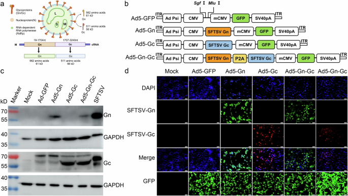
Referring to the M fragment of the genome sequence of the SFTSV JS2011-013-1 strain (GenBank NO. KC505127.1), the open reading frame (ORF) sequences of the SFTSV Gn (19-1704 bp) and Gc (1707-3240 bp) genes (Fig. 1a) were selected, cloned and inserted into the Ad5 vector by digestion with the restriction enzymes SgfI and MluI, followed by ligation with T4 DNA ligase as previously described39. The target sequences of SFTSV Gn, Gc, and Gn-Gc (linked by the P2A gene) were generated in the Ad5 vector (Fig. 1b). The recombinant adenovirus was constructed as previously described40. An adenovirus carrying the green fluorescence protein-encoding gene (Ad5-GFP) served as a control. Ad5-Gn, Ad5-Gc, and Ad5-Gn-Gc were propagated in HEK-293 cells, purified and stored at −80 °C. The insertion results were confirmed by PCR and sequencing. Viral titers (plaque-forming units/ml, PFU/ml) were determined in HEK-293 cells using the GFP-labeling method. HEK-293 cells were seeded in 96-well plates at a density of 2 × 10⁴ cells/well. The recombinant adenovirus samples were serially tenfold diluted from 10−1 to 10−8 with a final diluent volume of 400 μl. The culture medium of the HEK-293 cells in the 96-well plate was discarded, and 100 μl of each adenovirus dilution was added to the corresponding wells in triplicate. The cells were cultured for 24 h at 37 °C. The titers of adenovirus were observed and recorded under a fluorescence microscope. The number of fluorescent cells in the last two wells where fluorescence could be observed was recorded. The method for calculating the adenovirus titer was as follows: adenovirus titer (PFU/ml) = (A + B × 10) × 1000/2/(virus volume (μl) in A). A represents the average number of fluorescent cells in the last wells, and B represents the average number of fluorescent cells in the second to last wells.
Western blotting
The monolayers of HEK-293 cells were infected with recombinant adenovirus Ad5-Gn, Ad5-Gc, Ad5-Gn-Gc and Ad5-GFP (MOI = 10) or mock-infected with the same volume of DMEM for 72 h. Cells infected with SFTSV were used as a positive control. After infection, infected cells were lysed with RIPA buffer containing 1 mM PMSF (Beijing Solarbio Science & Technology Co., Ltd.). The lysates were centrifuged (16,000 rpm for 10 min) to remove precipitates. Equal amounts of protein were loaded onto a 10% SDS‒PAGE gel and transferred to PVDF membranes (0.2 μm, Millipore, Darmstadt, Germany). After blocking with 5% skim milk, the membranes were incubated with mouse anti-Gn monoclonal antibody (1:2000) or rabbit anti-Gc polyclonal antibody (1:1000) overnight at 4 °C. Subsequently, the membranes were incubated with HRP-conjugated anti-mouse or anti-rabbit IgG secondary antibodies (1:5000, Abcam, Cambridge, United Kingdom) for 1 h at room temperature. The positive signals were detected with enhanced chemiluminescence (ECL) substrate (Thermo, USA) with a Western Blotting Imaging System (General Electric Co., USA).
Immunofluorescence assay
The recombinant adenoviruses Ad5-Gn, Ad5-Gc, Ad5-Gn-Gc and Ad5-GFP or Mock inoculated with DMEM were inoculated for 48 h in Vero cells in 96-well plates (4 replicate wells for each group). Then, the cells were washed and fixed in precooled 80% acetone overnight at −20 °C. Mouse anti-Gn monoclonal antibody (donated by Professor Yu Xuejie from Wuhan University) and mouse anti-Gc polyclonal antibody (synthesized by Nanjing GenScript, China) were added to the corresponding wells, incubated at 37 °C for 1 h, and washed 3 times with PBS. Alexa Fluor 488-conjugated goat anti-mouse or Alexa Fluor 594-conjugated goat anti-mouse secondary antibodies (Abcam, Cambridge, United Kingdom) were added and incubated for 1 h, after which the nuclei were stained with DAPI and washed 3 times with PBS. Fluorescence microscopy (Olympus, IX71) was used to observe specific fluorescence.
50% tissue culture infective dose (TCID50) of SFTSV
Monolayers of Vero cells were inoculated with SFTSV. The virus culture medium was collected when the percentage of cell lesions exceeded 80%, and the supernatants were collected by centrifugation at 4 °C and 3500 rpm for 10 min and stored at −80 °C. The viruses were serially diluted tenfold and inoculated into Vero cells, which reached 80% confluence, in 96-well plates with 4 parallel controls. The 96-well plates were cultured at 37 °C in a 5% CO2 incubator for 3 d. Then, the culture media were discarded, and the cells were washed 3 times with PBS and incubated at 37 °C with rabbit anti-SFTSV Gn/Gc polyclonal antibody (1:400) followed by secondary goat anti-rabbit IgG H&L antibody (1:800). The results were observed and recorded under a fluorescence microscope. The viral titer (TCID50) was calculated using the Karber method: lgTCID50 = L + d(S-0.5)41. L represents the logarithm of the highest dilution, d represents the difference between the logarithms of the dilutions, and S represents the sum of the ratios of positive wells.
Immunization and pathogenicity evaluation
Six- to eight-week-old wild-type female BALB/c mice were randomly divided into 5 groups (n = 8 in each group). Then, 100 μl of DMEM or Ad5-GFP, Ad5-Gn, Ad5-Gc or Ad5-Gn-Gc virus dilutions (equivalent to 1 × 10⁸ PFU/ml) were injected intramuscularly (i.m.) into the corresponding mice. After immunization, the body weights and physical states of the mice were continuously monitored for 21 d.
Virus-neutralizing antibodies
At 2, 4, and 8 weeks after immunization, blood samples (n = 6 in each group at each time point) were collected to determine SFTSV virus neutralizing antibody (VNA) levels using a fluorescent reduction neutralization test (FRNT) as previously described22. Serum samples were separated and serially diluted two-fold with DMEM from 1:8 to 1:2048 and incubated with 100 TCID50 of the SFTSV JS-2011-013-1 strain for 1 h at 37 °C in a 5% CO2 incubator. When the Vero cells in the 96-well plate were 90% confluent, the above mixture of serum and virus or the control was added, and the cells were cultured for 48 h at 37 °C. After the cells were fixed with precooled 80% acetone, they were incubated with a mouse anti-Gc polyclonal antibody and then with an Alexa Fluor-488-labeled goat anti-mouse secondary antibody. The number of fluorescent spots was observed under a fluorescence microscope. When the number of fluorescent spots in the sample wells was reduced by 50% compared to that in the virus control wells, the corresponding serum dilution was considered the neutralizing antibody titer.
Flow cytometry
At 3, 6, and 9 d post-immunization (dpi), mice (n = 5 in each group at each time point) were sacrificed for humanitarian reasons, and single immunocyte suspensions of inguinal lymph nodes or peripheral blood were prepared and stained separately with antibodies (BD Biosciences, Franklin, TN, United States) against DCs (CD80+, MHC I+, MHC II+, CD86 and CD11c+) or B cells (CD40+ and CD19+). The flow cytometry procedure was performed with a BD FACSCelesta flow cytometer, and the data were analyzed using FlowJo software.
Enzyme-Linked Immune Spot Assay (ELISpot)
At 28 dpi, the spleens of the immunized mice (n = 6 in each group at each time point) were collected. Lymphocytes in the spleen were dissociated and stimulated with 2 μg of purified SFTSV or Gn protein for 20 h in a 37 °C incubator with 5% CO2. Mouse IFN-γ/IL-4 ELISpot kits (Dakewe Biotech Co. Ltd., China) were used to determine the number of lymphocytes that secreted IFN-γ or IL-4 according to the manufacturer’s instructions. The number of spot-forming cells (SFCs) was calculated by eliminating background responses with an ELISpot plate reader (AID‘ ELISPOT reader-iSpot, AID GmbH, Germany).
Serum-specific IgG subtypes
IgG typing data were identified by ELISA. At 2, 4, and 8 weeks after immunization, the serum levels of IgG1 and IgG2a antibodies against SFTSV in the Ad5-Gn, Ad5-Gc and Ad5-Gn-Gc groups were detected. A total of 600 ng of intact SFTSV protein per well was precoated in 96-well plates, and the titers of IgG1 and IgG2a were detected by ELISA, ensuring that the antibody levels in the serum of immunized mice specific for SFTSV Gn or Gc could be detected. After washing and blocking, 100 μl of 10-fold serially diluted sera from the Ad5-Gn, Ad-Gc and Ad5-Gn-Gc groups were added and incubated overnight at 4 °C. The plates were washed and then incubated at room temperature for 3 h with 100 μl of HRP-conjugated goat anti-mouse IgG1 and IgG2a at a dilution of 1:500. ABTS was added, and the plates were incubated at 37 °C for 15 min after washing. The absorbance at 405 nm was measured using a microplate reader (Mutishan MK3, Thermo Scientific). Serum samples from mock-immunized mice were used as negative controls. Sample titers were considered to be positive if the absorbance reading was at least 2.1 times that of the mock group. The positive antibody end-point titers are expressed as the reciprocals of the highest dilution of serum42.
Mice IFNAR Ab treatment and virus challenge
Six- to eight-week-old female BALB/c mice were randomly divided into 5 groups (n = 8 in each group). The immunization schedule is shown in Fig. 6a. 100 μL of DMEM, Ad5-GFP, Ad5-Gn, Ad5-Gc or Ad5-Gn-Gc virus dilution (equivalent to 1 × 10⁸ PFU/ml) was injected i.m. into the corresponding mice. At 27 dpi, a MAR1-5A3 (mouse anti-mouse IFNAR, IgG1) monoclonal antibody (mAb) (Leinco Technologies, United States) was intraperitoneally (i.p.) injected at a dose of 1.25 mg per mouse 1 d before SFTSV infection to block type I IFN signaling19. At 28 dpi, IFNAR Ab-treated mice were challenged with SFTSV at a dose of 1 × 105.25TCID50. Body weights and clinical manifestations were continuously recorded for 21 d after the virus challenge. We established a scoring standard based on diet, sleep, behavior, body weight, and hair to evaluate the health status of infected mice in detail (Table 1). Total RNA from the spleen, liver, lung, and brain tissues of IFNAR Ab mice on the 7th d post-SFTSV challenge was collected with an RNAprep Pure Tissue Kit (TIANGEN Biotech, Beijing Co. Ltd., China) for real-time PCR detection (DaAnGene Co. Ltd., China) of SFTSV viral loads following the manufacturer’s instructions.
Table 1 Clinical symptom scoring standard
Histopathology
The spleens and livers of IFNAR Ab-treated mice were collected on the 21st d or before death after the SFTSV challenge and fixed with 4% paraformaldehyde for 24 hours. After gradient alcohol dehydration and clearing, the tissues were paraffin-embedded and sectioned into 5 μm slices. The slices were stained with hematoxylin and eosin for histological observation.
Passive transfer of serum from vaccinated mice to IFNAR Ab-immunized mice
Wild-type 6- to 8-week-old female BALB/c mice (n = 6 in each group) were injected with the MAR1-5A3 mAb 24 h prior to SFTSV infection, as described in section “Mice IFNAR Ab Treatment and Virus Challenge”. The immunization schedule is shown in Fig. 8a. Twelve hours following the injection of the MAR1-5A3 mAb, naive IFNAR Ab mice were injected intraperitoneally with 200 μl of serum specific for SFTSV Gn, high-titer (VNA titers > 1:250) or low-titer (VNA titers ≤1:250) serum specific for SFTSV Gc, or DMEM. IFNAR Ab mice with passive transfer of serum were challenged with 1 × 105.25 TCID50 of SFTSV. Afterwards, body weights and clinical manifestations were monitored for 21 d.
Statistical analysis
The experiments were repeated, and the data were collected at least 3 times. The data were analyzed using SPSS 26.0 statistical software (SPSS Inc., Chicago, IL, United States) and are presented as the mean ± standard deviation. Statistical differences among the groups were identified through one-way ANOVA. P < 0.05 was considered to indicate statistical significance. Graphs were drawn with GraphPad Prism 8 statistical software. *p < 0.05; ** p < 0.01; *** p < 0.001.

